V7 Darwin
Healthcare AI · Medical Imaging
Overview
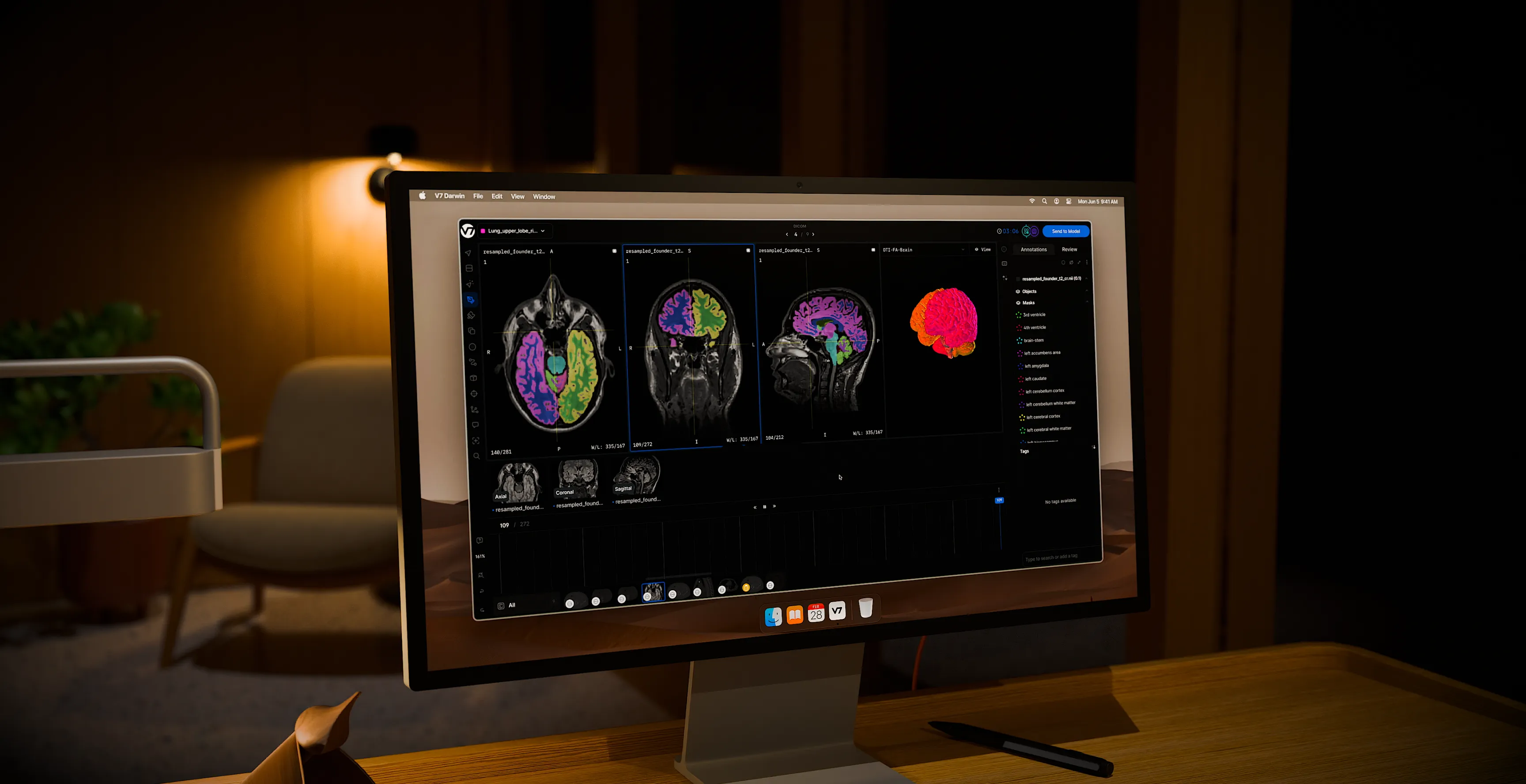
I designed a specialized medical-imaging workspace that lets healthcare AI teams annotate, review, and export complex clinical data with diagnostic precision, collapsing a fragmented, error-prone toolkit into a single traceable pipeline.
Role
Product Designer, lead on workspace and workflow integration
Domain
Healthcare AI, medical imaging, enterprise SaaS
Primary users
Radiologists, pathologists, ML engineers, and data-operations teams
The workspace is where medical AI teams build their training data: they load volumetric scans, label anatomy with AI assistance, run expert review, and export clean, structured datasets into a model pipeline. Everything in this case study sits inside that one dense diagnostic canvas.
The problem
Before this workspace existed, medical AI teams ran their pipeline across four disconnected tools. Radiologists annotated in local DICOM and NIfTI viewers, logged findings into spreadsheets, managed task assignments through internal scripts, and discussed cases in a separate messaging app.
That fragmentation broke down on three fronts:
For clinicians
Context evaporated at every tool switch. The diagnostic reasoning behind a label lived in someone’s head or a spreadsheet cell, not next to the image.
For reviewers
There was no traceability. They could see what was labeled but never why, which made disagreement impossible to resolve cleanly.
For the business
The manual handoffs slowed dataset development, inflated QA overhead, and created compliance exposure in a regulated environment where every annotation may need an audit trail.
The design pivot
The instinct with complex enterprise tools is to simplify, to hide density behind clean surfaces. For expert clinical users, that is wrong. The goal was not to make medical imaging look simple. It was to take dense, high-stakes data and organize it so it became scalable, traceable, and AI-ready. Structure the complexity, do not mask it.

PROCESS
Ingest → Dataset org and metadata → Annotation and AI assist → QA and consensus review → Pipeline export
Discovery: clinical habits vs. ML requirements
I started by mapping two sets of needs that rarely meet in one tool. Clinicians need familiarity: windowing, crosshairs, oblique planes, multi-planar reconstruction, slice timelines. ML teams need structure: error-free schemas, clean JSON exports, complete audit logs. The workspace had to serve both without forcing either group to compromise.
Two principles that drove the design
Familiarity first, intelligence second
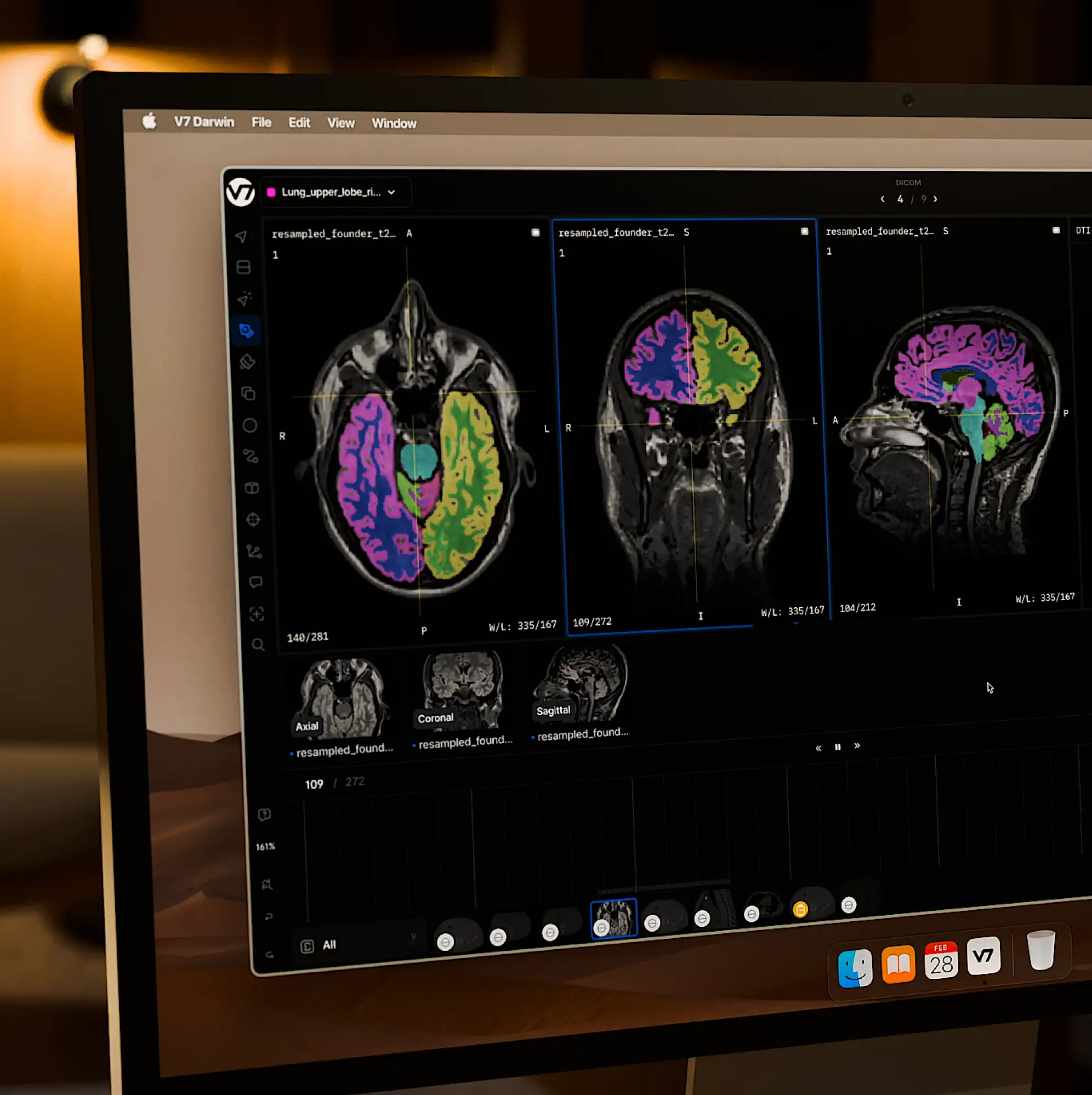
I anchored the canvas in patterns radiologists already trust, DICOM-compliant viewing, multi-slot layouts, multi-channel imaging, then layered intelligence on top: inline AI segmentation and consensus status. The AI never displaces the clinician’s mental model, it augments it.
Organize density, do not hide it
Deep controls stay accessible but ordered, collapsible context-aware inspector docks instead of buried menus, so the canvas keeps maximum real estate without the tooling ever going out of reach.
MY ROLE
As the sole product designer on the core workspace, I owned the end-to-end interaction models, panel architecture, and tool modalities for the diagnostic canvas. Working with the PM, engineers, and clinical advisors, my hands-on contributions were:
Layout geometry and information architecture
Prioritizing viewport real estate for the multi-slice canvas and organizing deep parameters into collapsible, context-aware inspector docks.
Multi-planar reconstruction (MPR)
Prototyping the full MPR configuration: mouse-drag behavior, active-axis crosshair sync, and slice-scroll physics.
Component taxonomy for dense UI
Reusable patterns for layer toggles, multi-channel inputs, and contextual AI prompts, integrated into the core design system.
Clinical alignment
Running critique sessions with radiologist advisors to validate windowing and level adjustments before each engineering sprint.
Key decisions
DICOM-compliant core workview
Preserves native viewing patterns: windowing, crosshairs, MPR.
Outcome: removes the learning curve for radiologists and protects diagnostic trust from day one.
Inline, in-cursor AI tooling
Segmentation models (MedSAM, TotalSegmentator) live in the active toolkit, not a background batch job.
Outcome: speeds the active labeling cycle while keeping the expert in control to correct AI output immediately.
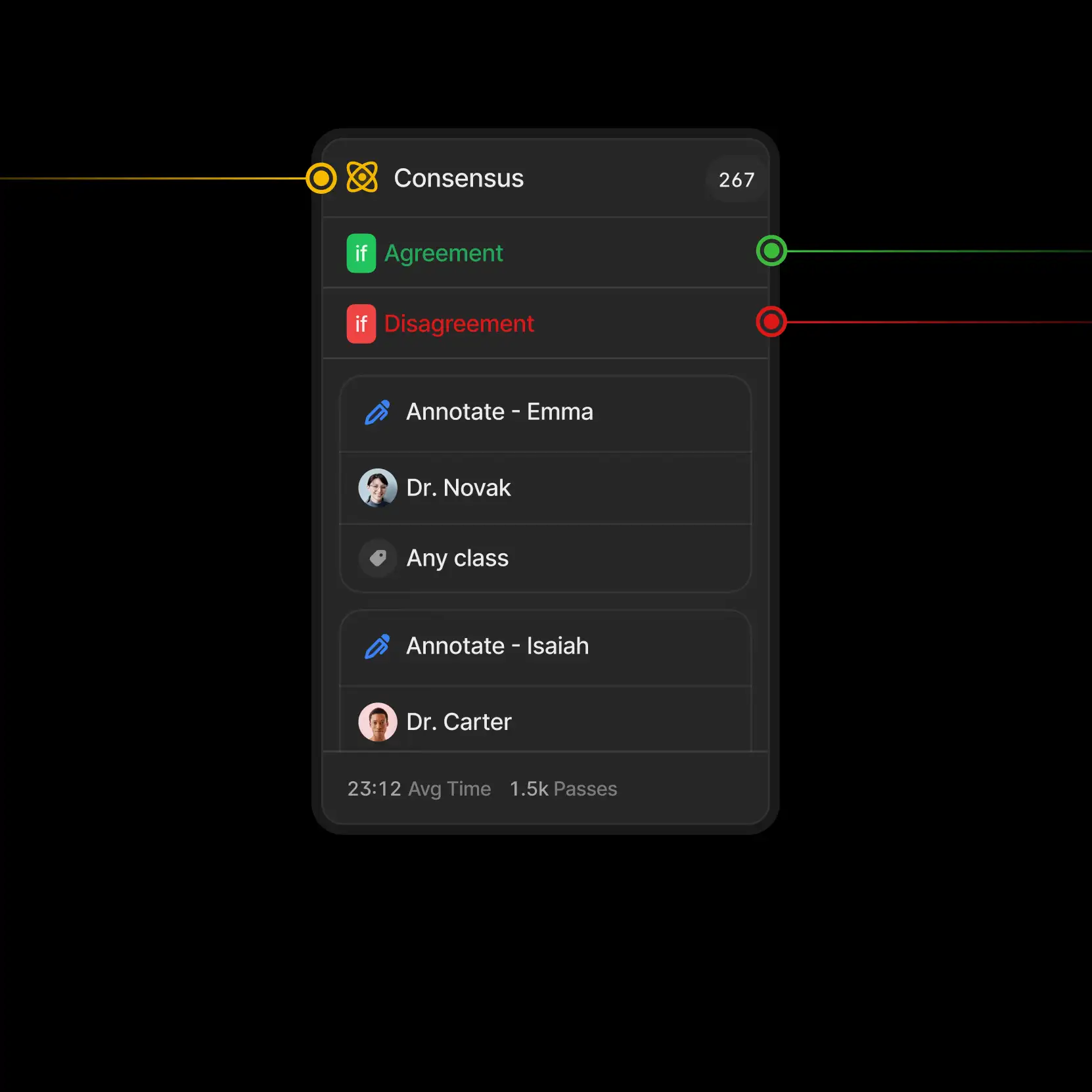
Traceable QA consensus engine
Side-by-side blind reads with inline dispute comments and task-state management.
Outcome: raises dataset quality by surfacing exactly where expert opinions diverge, and why.
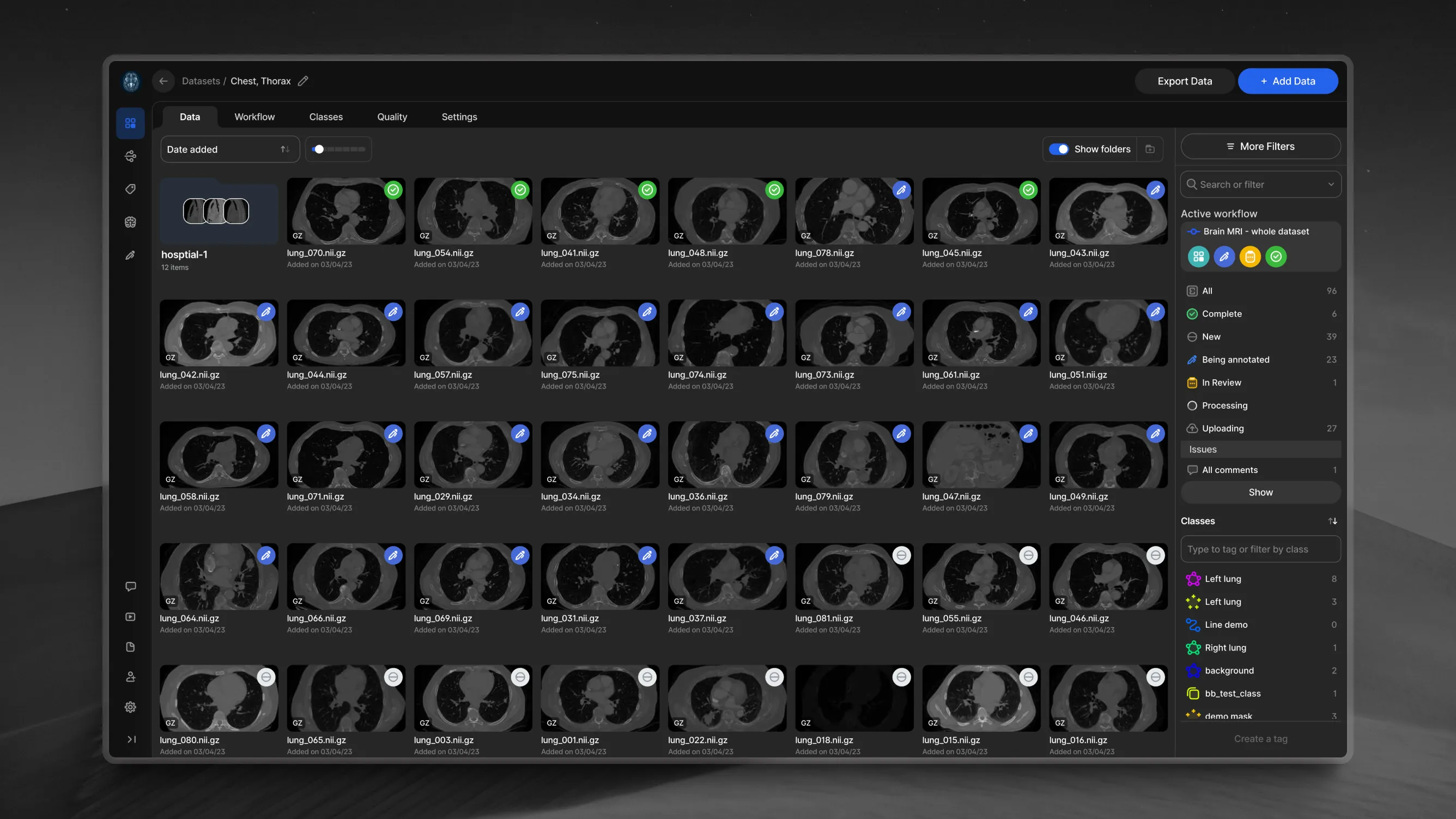
Pipeline-native data architecture
Metadata tagging, folder organization, and direct exports designed alongside the canvas, not bolted on.
Outcome: ML engineers pull clean, structured datasets with no manual reformatting.
Experience & craft
The diagnostic workview
A dense, high-contrast dark-mode workspace built for long viewing sessions. Advanced controls, layers, multi-slot view adjustments, multi-channel inputs, sit in persistent panels so clinicians never lose sight of the canvas.
Human-in-the-loop AI
Tools like Auto-Annotate and Flood Fill render real-time vector previews. Users refine AI boundaries with brush modifiers before committing a label, so the expert always has the final say and errors get caught at the source.
Edge cases and safeguards
Most of the design work that mattered lived in the failure states:
Multi-gigabyte load latency
Volumetric CT and MRI files routinely caused rendering lag and input freezes. With engineering, I designed asynchronous loading states, localized segment-caching indicators and a read-only canvas overlay, that keep users informed without breaking flow or risking a crash.
Lost work on network drops
A user could spend 15 minutes refining a contour and lose the unsaved vector to a brief disconnect. I designed an aggressive local-storage delta auto-save plus an offline banner that safely pauses interaction when the connection degrades.
Regulated audit trails
For training data to stay legally viable, every cursor modification has to be logged. I designed a background metadata tracker that captures every adjustment without cluttering the viewport with status noise.
VALIDATION
I ran task-based testing with practicing radiologists and clinical research coordinators.
The task
Load an unfamiliar multi-slice NIfTI volume, use MedSAM to isolate a specific anatomical feature, correct an intentionally planted boundary error, and submit to a blind-review workflow.
The key finding
Early versions hid layer visibility behind a dropdown, and users repeatedly lost track of which slice plane they were editing, a spatial-awareness failure that is dangerous in a diagnostic tool. I refactored to a persistent structural panel that anchors plane awareness at all times, and the confusion disappeared in follow-up sessions.
REFLECTIONS
Complexity is a feature
For expert-facing tools, hiding density behind minimalism adds friction rather than removing it. Utility comes from organizing complexity so it stays legible and traceable, not from masking it.
What is next
I would optimize the onboarding loop for pathology-specific workflows like whole-slide imaging, and add cross-slice keyboard shortcuts to cut manual manipulation during high-volume segmentation.
Let's talk
Pick whatever works best for you, a quick video call or a simple email.